攝護腺類肉瘤癌—案例報告
潘柏勳1、林子平1,2、黃志賢1,2
1台北榮民總醫院 泌尿部;2國立陽明大學醫學院泌尿學科 書田泌尿科學研究中心
Prostate sarcomatoid carcinoma with chondro-osseous differentiation: a Case Report
Po-Hsun Pan1, Tzu-Ping Lin1,2, William J. Huang1,2
1Department of Urology, Taipei Veterans General Hospital, Taipei, Taiwan
2 Department of Urology, School of Medicine and Shu-Tien Urological Institute,
National Yang-Ming University, Taipei, Taiwan
Introduction:
Prostate sarcomatoid carcinoma is an extremely rare and aggressive malignant disease which caused poor prognosis. This very little subtype just accounts less than one percent of all prostate cancer. Some of these cancers may not have PSA elevation which could increase the difficulty of detection and monitoring. Here we presented one case of sarcomatoid carcinoma of prostate and the following management.
Case report:
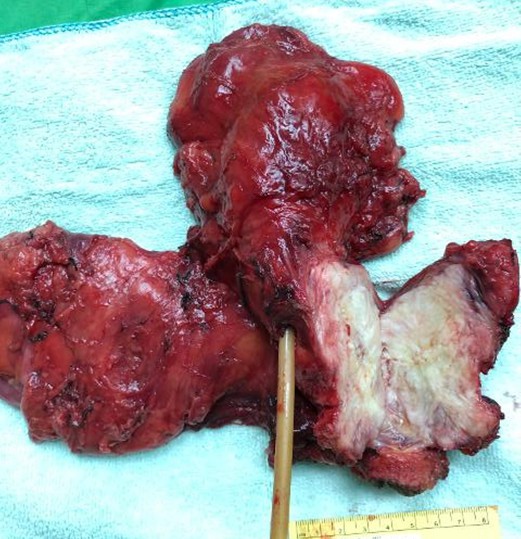
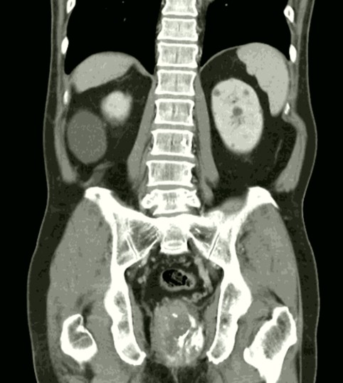
A 75-year-old man has history of hypertension under medical treatment but no operation history. Initially, the patient presented with lower urinary tract symptoms, including frequency, urgency, nocturia 3 times per night, weak stream, intermittency, straining and incomplete emptying for three months. Anal pain was noted with tenesmus after then. So, he first presented to colorectal surgery department for survey where digital rectal examination showed an extraluminal mass prolapse from anterior wall of rectum. Tumor markers were checked which showed elevated PSA up to 72.50 ng/ml, with free PSA to PSA ratio of 0.08. On the other hand, CEA was in normal range (1.3 ng/ml, normal < 5 ng/ml). Hence, he then referred for further survey of prostate cancer. Transrectal ultrasound guided prostate biopsy was performed and the pathology revealed malignant tumor with chondro-osseous differentiation. Colonoscopy showed the tumor was grossly infiltrated about 3cm in size and located at 2cm from anal verge. Imaging survey of pelvis CT revealed an irregular mass about 9.9 x 5 x 3.6cm in size with heterogeneous internal enhancement and calcifications at low pelvis with protruding to posterior wall of urinary bladder and external compression at rectum. Tumor involved to bladder and rectal wall was suspected. Besides, an enlarged lymph node near left external iliac vessel was also noted. So, the impression was osteogenic sarcoma of prostate, cT4N1M0. He received radical prostatectomy combined with radical cystectomy, abdominal perineal resection, colostomy and ileal conduit reconstruction. The operation was done smoothly and the patient tolerated well. The patient recovered well post-operatively. Pathology reported sarcomatoid carcinoma with bladder, rectum and pelvis lymph node invasion, pT4N1M0. Androgen deprivation therapy was prescribed one month post-operatively. Local radial therapy was also applied to surgical bed and lymph node. Further follow up and adjuvant therapy will be arranged.
Conclusion:
Prostate sarcomatoid carcinoma is a rare malignant disease of prostate. In previous case reports and series, it is hard for diagnosis due to the absence of PSA raising. It is also difficult for treatment owing to the poor response to hormone therapy, chemotherapy and radiotherapy. However, definitive treatment of en-bloc resection with other adjuvant therapy might give a satisfactory treatment effect. Further novel therapy such as immunotherapy may also give a role in such disease treatment.
附件: