偽裝成後腹腔腫瘤之靜脈血管畸形-個案報告
邱俊凱、李建儀1、江奕2
台中榮總外科部泌尿科;台中榮總病理部
Venous malformation mimicking Retroperitoneal malignant tumor – A Case report.
Chiou JK, Li JR1, Chiang I2
Divisions of Urology, Department of Surgery, Taichung Veteran General Hospital, Taichung, Taiwan, Department of Pathology, Taichung Veteran General Hospital, Taichung, Taiwan
Abstract:
Vascular malformation is rare and results from errors of vascular morphogenesis. Malformations of venous origin are usually asymptomatic. Their location into the retroperitoneum is extremely rare. Only very few cases of retroperitoneal vascular malformations are available in literature. Herein we report a case of venous malformation with entrapment of left renal hilum vessels presenting as a retroperitoneal mass, managed successfully by surgical excision.
Introduction:
Retroperitoneal cystic masses can be classified as neoplastic and nonneoplastic. Neoplastic lesions usually contain irregular cystic walls with heterogeneous features while Nonneoplastic ones are always smooth and homogenous. CT scan is the major tool for differential diagnosis. Surgical interventions are required for neoplastic lesions and symptomatic nonneoplastic ones. For some cases, image study cannot completely verify, Clinical dilemma wound develop. We presented a case of symptomatic retroperitoneal cystic mass which had difficulty in pre-operative differential diagnosis.
Case report:
52-year-old women presented to surgical emergency with left lower quadrant abdominal dull pain intermittently for one week. The pain exaggerated combined with decrease of appetite. She had history of Myomectomy 16 years ago, but there was no feature suggestive of intestinal obstruction.
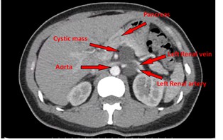
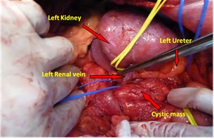
On examination, the patient was non-febrile. There was no knocking pain nor abdominal tenderness while palpation except a mass lesion found at left upper abdomen. Lab data also showed no infection nor inflammation sign. Abdominal CT scan revealed a 9cm cystic lesion located beneath pancreatic body with entrapment of left renal hilum. The cystic fluid was homogenous without wall thickening and the region of interest of the cyst was 7 Hounsfield units. She received retroperitoneal exploration under the impression of cystic retroperitoneal tumor, r/o malignancy. During operation, a soft, well capsulated cystic lesion was found in between aorta and kidney, incasing the renal vessels. One supplying vessel originated from aorta between the left renal artery and inferior mesentery artery root was identified. After meticulous dissection of the adjacent adhesive tissues, the mass was excised without scarification of left renal tissues. Histopathological showed a cystic lesion, which is consisted of irregularly thickened fibromuscular layers and lined by endothelial cells, confirmed to be a venous malformation. The patient encountered chyle leakage from drainage tube on the second post-operative day. After fat free diet, the symptom improved and she was discharged on seventh post-operative day.
Discussion:
This is the first adult case of retroperitoneal venous malformation extending between pancreas, aorta, and renal vessels to be reported. Vascular malformations are benign vascular abnormal growths that usually present in young ages. They do not cause any health problems but occasionally interfere with several functions of the body depending on their location. The abnormal growths of blood vessels may occur in the muscles, visceral organs and more commonly on skin surface. These lesions can classified into several different types including capillary, venous, arterial, lymphatic and combine. Majority of venous malformations did not draw our attention until they are first observed. Furthermore, more than 90% disappear spontaneously when the patient reaches puberty.
Retroperitoneal venous malformation is a very rare condition. It’s a result of angioblastic tissues malformation during fetal life. There are only several cases reported so far. Precise pre- and intra-operative diagnosis of retroperitoneal venous malformation is difficult. The lesion is usually detected only if it had clinical symptoms which caused by the mass pressure to the surrounding tissues. There are some retroperitoneal vascular malformation mimicking different disease had been reported earlier.
The clinical implications and therapeutic strategies for retroperitoneal cystic masses vary depending on the cause. Although CT scan is essential for differential diagnosis, there are still substantial overlap of image findings in various retroperitoneal cysts. For this patient, the feeding artery of the cyst was not identified in CT scan. In the future, CT angiography should be considered in retroperitoneal masses which are close to major vessels.
In this patient, pre-operative CT scan showed bizarre appearance. It contained homogenous benign feature and lobulated structures with renal vessels entrapment which implied malignant potential. We decided to dissect the tumor with possibility of en bloc wide excision. However, during operation, the cystic wall was smooth and nonadhesive. Benign entity was highly suspicious and the kidney was preserved. If we have venous malformation as our pre-operative differential diagnosis, the surgical decision would be easier to make.
附件: