後腹膜腔平滑肌肉瘤源於腎臟筋膜經腹腔鏡腎臟保留手術切除之預後
許哲維1、張彰琦1,3、邱逸淳2,3、洪士奇1
1台北市立聯合醫院忠孝院區外科部泌尿科;
2台北市立聯合醫院和平婦幼院區外科部泌尿科;
3 國立陽明大學醫學院泌尿學科
2台北市立聯合醫院和平婦幼院區外科部泌尿科;
3 國立陽明大學醫學院泌尿學科
Retroperitoneal Leiomyosarcoma Growing from Gerota’s Fascia:
Laparoscopic Renal-sparing Surgery Surgical Outcome and Follow-up
Laparoscopic Renal-sparing Surgery Surgical Outcome and Follow-up
Che-Wei Hsu1, Chang-Chi Chang1,3, Yi-Chun Chiu2,3, Shih-Chi Hong1
1Division of Urology, Department of Surgery, Zhongxiao Branch, Taipei City Hospital, Taiwan
2 Division of Urology, Department of Surgery, Heping-Fuyou Branch, Taipei City Hospital, Taiwan
3 Department of Urology, School of Medicine, National Yang-Ming University, Taiwan
Introduction: Leiomyosarcoma (LMS) is a rare type of cancer, accounts for less than 1% of all connective tissue malignancies. Retroperitoneal leiomyosarcoma is predominantly observed in elderly women and mostly found in the abdominal cavity and pelvic regions. Herein we present an 80-year-old man with retroperitoneal leiomyosarcoma who received laparoscopic renal-sparing surgical resection followed by radiotherapy.
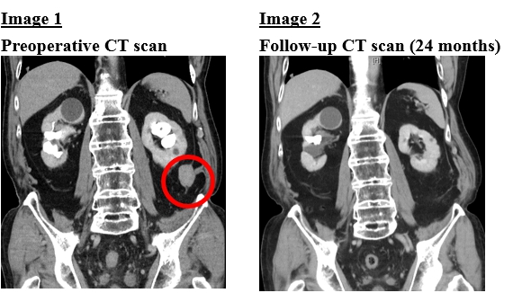
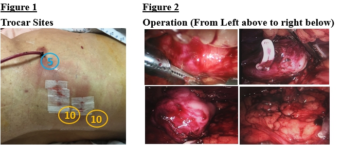
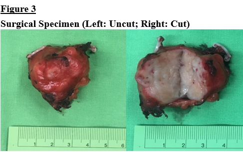
Case Report: This 80-year-old male presented with left flank pain. He had history of urolithiasis many times. CT scan of abdomen showed left ureteral stone with obstructive uropathy and one 3.2 cm lobulated enhanced soft tissue mass lesion at left lower gerota fascia (Image 1). He decided to have ureteroscopic lithotripsy and ultrasound-guided tumor biopsy, which revealed tumor composed of the neoplastic cells with atypia presenting focal obvious pleomorphism. Further surgical intervention was suggested. He received left retroperitoneal laparoscopic-assisted renal-sparing tumor resection. Under general anesthesia, the patient was positioned as modified right decubitus position. Two 10mm trocar sites were created at left posterior axillary line. One was above iliac crest and the other was below the 12th rib. One 5mm trocar site was created at mid-axillary line and above iliac crest (Figure 1). The tumor border was resected from gerota’s fascia and the tumor base was electro-coagulated without injury to renal parenchyma (Figure 2). Total operative time was 130 minutes and the blood loss was 110 ml. Pathological report was leiomyosarcoma, grade 1 (FNCLCC system) (4.0 x 3.0 x 2.2 cm) (Figure 3). The patient was conducted adjuvant radiotherapy. Follow-up CT scan at 24 months after the operation, showed no remarkable discrete enhanced soft tissue mass is seen in at left flank region (Image 2).
Conclusion: About 10%-37% of all soft tissue sarcomas arise in the retroperitoneum. Surgical resection is the main treatment for retroperitoneal leiomyosarcoma and improves long-term prognosis. Adjuvant radiotherapy is associated with a reduced risk of local recurrence. We present the case of retroperitoneal leiomyosarcoma growing from gerota’s fascia receiving laparoscopic-assisted renal-sparing tumor resection followed by radiotherapy. He is currently alive without recurrence or metastasis. From this experience, once incidental retroperitoneal tumor is noted, leiomyosarcoma might be take in to consideration of differential diagnosis and renal-sparing tumor resection followed by radiotherapy could be one of the treatment options.
附件: