不同亞型婦女反覆膀胱發炎之膀胱表皮細胞增生、細胞骨架、及屏障蛋白質之表現
李秉叡、江元宏、郭漢崇
佛教慈濟醫療財團法人花蓮慈濟醫院 泌尿部
Urothelial cell proliferation, cytoskeleton and barrier function protein expression in the female patients with different phenotypes of urinary tract infection
Ping-Jui Lee, Yuan-Hong Jiang, Hann-Chorng Kuo
Department of Urology, Hualien Tzu Chi Hospital, Buddhist Tzu Chi Medical Foundation and Tzu Chi University, Hualien, Taiwan
Purpose: Chronic inflammation, urothelial cell apoptosis and impairment of barrier function could be the underlying pathophysiology of recurrent urinary tract infection (UTI) in women. The present study aims to investigate the difference of inflammatory cell, apoptotic proteins, regenerative cytokines, barrier function proteins, and sensory proteins expression between recurrent bacterial cystitis and normal population in women.
Materials and Methods: This retrospective, single-center study was performed in Hualien Tzu Chi Hospital (Hualien, Taiwan), and approved by the Institutional Review Board and Ethics Committee of the hospital. The written informed consent was obtained after informed the study rationale to each patient. The patients recruited in this study consisted of women at least 20 years old who had experienced at least two episodes of symptomatic UTI within the past 6 months or three episodes of symptomatic UTI within the past year. They were further stratified into persistent bacteriuria and non-persistent bacteriuria based on the severity of UTI. For comparative purposes, a group of women who were free from recurrent UTI were chosen as a control group. The bladder mucosa using a cold-cup biopsy were taken and investigated. The expressions of inflammatory biomarkers (Tryptase, P38), apoptotic proteins (BAD, BAX, Caspase-3), regenerative cytokines (CK5, CK14, CK20, SHH, TP63, CD34), barrier proteins (E-cadherin, zonula occludens-1), muscarinic receptors (M2, M3), and adrenergic receptor (β3) were compared by Western blotting with quantification between the recurrent UTI group and the control group. Hematoxylin and eosin (H&E) staining and Immunofluorescence staining (IHC) with qualification for inflammatory biomarker (Tryptase), barrier proteins (uroplakin III), proliferative protein (Ki67) and Terminal deoxynucleotidyl transferase dUTP nick end labeling (TUNEL) assay were also performed in the bladder specimens.
Results: A total of 21 women with recurrent UTI and 7 control group participants without recurrent UTIs were included in this study. The mean ages of the two groups were 68.3 ± 9.0 years and 64.3± 9.9 years, respectively. The quantification of western blot analyses showed significantly higher Caspase-3, CK5, and CK14 expression (P=0.008, P=0.002, and P<0.001, respectively), whereas lower P38 and CK20 expression (P<0.001 and P=0.031, respectively) in the recurrent UTI patients when compared with the control group. Further analyses were performed for the subgroups of patients with persistent and non-persistent bacteriuria. The inflammatory protein, P38, was significantly higher in the control group, the apoptotic proteins as BAX and Caspase-3 were significantly higher in subgroup of persistent bacteriuria, and proliferative protein, CK14, was significantly higher in subgroup of non-persistent bacteriuria. It showed a trend that barrier proteins were lower in both persistent and non-persistent bacteriuria groups while the difference were not significant. [Figure 1] In H&E and IHC qualitative analyses, the expression of UPK3 were weaker and disarranged in persistent and non-persistent UTI patients than the controls. The apoptotic cells stained with TUNEL were less observed in the control group [Figure 2].
Conclusions: The increased urothelial cell apoptotic, proliferative, and decreased barrier function proteins expression were observed in the patients with recurrent UTI than in the control group.
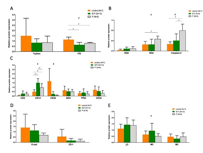
Figure 1. The Western blotting with quantification of urothelial cell proteins in the patients with recurrent UTI group and the control group. (A) Inflammatory proteins, (B) Apoptotic proteins, (C) Proliferative proteins, (D) Barrier proteins, and (E) Autonomic receptor proteins.
Abbreviations: P, Persisted bacteriuria; N-P, Non-persisted bacteriuria;
# Indicates significant difference, P value < 0.05, in ANOVA analysis
* Indicates significant difference, P value < 0.05, in post-hoc analysis
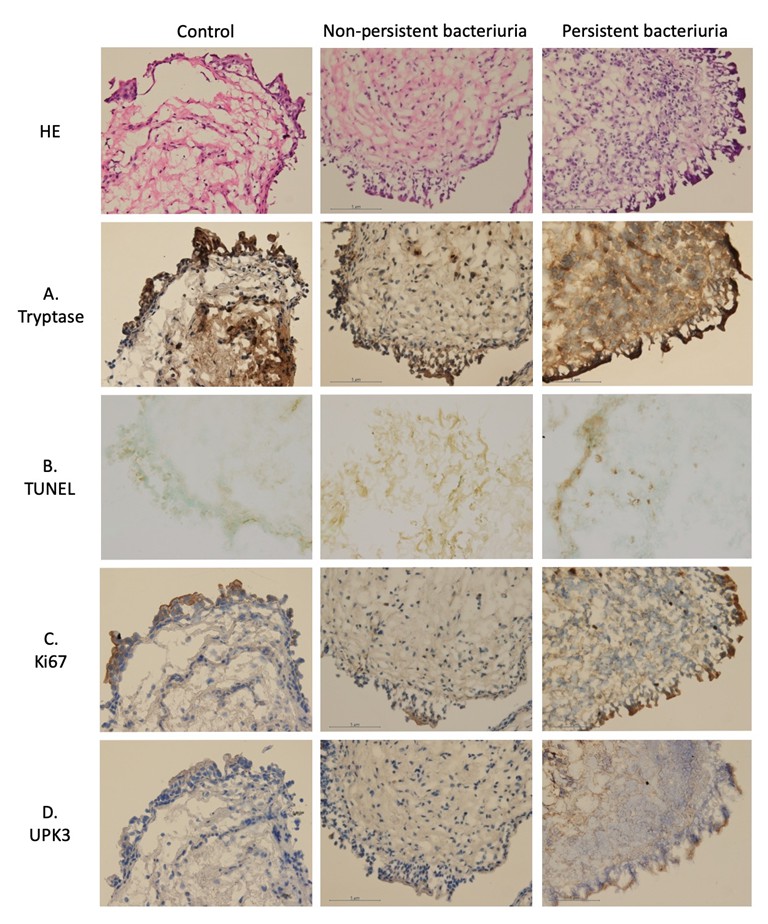
Figure 2. The Western blotting with quantification of urothelial cell proteins in the patients with recurrent UTI group and the control group. (A) Inflammatory proteins, (B) Apoptotic proteins, (C) Proliferative proteins, (D) Barrier proteins
附件: