嗜酸性粒細胞瘤與腎臟細胞瘤之影像鑑別
朱信誠、何承勳
台北醫學大學-部立雙和醫院泌尿科
Case Report-Discrimination of Oncocytoma and Renal Cell Carcinoma
Chu-Hsin Cheng, Chen-Hsun Ho
Department of Urology, Taipei Medical University-Shuang Ho Hospital, Taipei, Taiwan
Backgroud:
Renal cell carcinoma (RCC) is the second most common primary tumor of the urinary system. Current literature reports that oncocytoma represents 2–11% of all renal tumors[1-3]. Oncocytoma is typically described to be a hypervascular, homogenous mass that may contain a central stellate scar on computerized tomography (CT). However, Oncocytoma and RCC remains difficult to discriminate from the CT image. Therefore, we present a case that was diagnosed with Oncocytoma by radiologist pre-operatively, which was proven to be clear cell RCC pathologically.
Case Report:
This 71-year-old woman has pasted history of type 2 diabetes mellitus and hypertension under medication control in Republic of Marshall Islands. She was presented with left sided kidney mass on CT scan accidentally a month ago (Figure 1.). The tumor was 5*4*4 cm with radial scar at upper pole of left kidney, suspect oncocytoma, reported by radiologist.
There was no palpable mass, gross hematuria, flank pain, or body weight loss. Left side Laparoscopic partial nephrectomy was performed, and warm ischemia time was 14 min 00 sec. Surgical histopathology showed clear cell renal cell carcinoma. Creatinine level was 1.04 mg/dL before the surgery, and increased to 2.02 mg/dL on post-operative day 1. After adequate hydration, creatinine level decreased to 1.02 mg/dL. There was no severe complication, and the patient was discharged after a week.
Conclusion:
In this case, the mass was well defined, with regular margin and central stellar scaring. Oncocytoma was diagnosed by radiologist. However, malignant tumor such as RCC, should also be considered, given the size of the mass. Previous literature showed that renal mass biopsy was found to be unreliable in confidently diagnosing a localized renal mass as an oncocytoma with one in four found to be RCC on surgical pathology [4] (Figure 2). Complete resection should be an treatment option for renal tumor despite the benign picture radiologically.
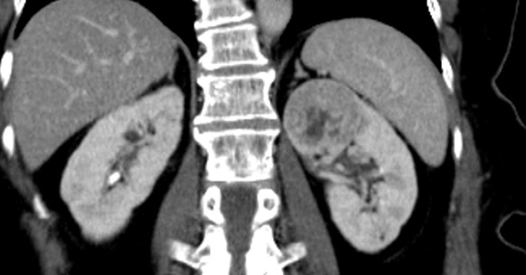
Figure1. A 5*4*4 cm tumor with radial scar at upper pole of left kidney.
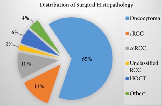
Figure2.
Surgical histopathology results for all suspected oncocytomas in the systematic review. HOCT, hybrid oncocytic/chromophobe tumor; ccRCC, clear cell RCC; pRCC, papillary RCC; cRCC, chromophobe RCC.
Reference:
1. Pradhan D, Kakkar N, Bal A, Singh SK, Joshi K (2009) Subtyping of renal cell tumours; contribution of ancillary techniques. Diagn Pathol 4:21
2. Rekha PR, Rajendiran S, Rao S, Shroff S, Joseph LD, Prathiba D (2008) Histological reclassification, histochemical characterization and c-kit immunoexpression in renal cell carcinoma. Indian J Urol 24(3):343–347
3. Snyder ME, Bach A, Kattan MW, Raj GV, Reuter VE, Russo P (2006) Incidence of benign lesions for clinically localized renal masses smaller than 7 cm in radiological diameter: influence of
sex. J Urol 176(6 Pt 1):2391–2395
4. BJU Int. 2017 May;119(5):661-666. doi: 10.1111/bju.13763. Epub 2017 Feb 27.
附件: