Podium 01
病例報告: 原發性腎臟肌周皮細胞瘤
李懿文1、劉惠瑛1、宋明澤2
1高雄長庚紀念醫院泌尿科、2高雄長庚紀念醫院病理科
Cases Report: Primary kidney myopericytoma
I-Wen Lee1, Hui Ying Liu1, Ming-Tse Sung2
1Department of Urology, Chang Gung Memorial Hospital, 2Department of Pathology, Chang Gung Memorial Hospital, Kaohsiung Medical Center, Kaohsiung, Taiwan.
[Introduction]
Myopericytomas are unusual benign perivascular neoplasms typically originating from the skin and superficial soft tissues of distal extremities, trunk, and head and neck regions. These tumors show a perivascular concentric multilayering of ovoid- to spindle-shaped cells with a myoid appearance. They are extremely rare in visceral locations and only few cases of renal myopericytoma had been published. Because of its rarity, the morphologic features, immunohistochemical profiles, and biological behavior of this tumor have not been entirely understood. Thus, we present a case of renal myopericytoma status post partial nephrectomy which might be the first case of renal myopericytoma reported in Taiwan.
[Case history]
A 71-year-old Asian male with a past medical history of hypertension, type 2 Diabetes Mellitus, liver hemangioma and tongue squamous cell carcinoma status post radical surgery on January,2022. Right renal 4 cm iso-echoic nodule was accidentally found by abdominal echography during regular follow-up of liver hemangioma. The patient was referred to the Outpatient Department (OPD) of Nephrology in our hospital under the first impression of right kidney tumor.
The physical examination and urine analysis did not show further positive findings. Renal echography showed right renal 4 cm iso-echoic intraparenchymal mass. Subsequent enhanced Computed Tomography of abdomen discovered a 4.8 cm right renal heterogenous enhancing mass without definite enlargement of retroperitoneal lymph nodes. (Fig.1,2). The preoperative RENAL Nephrometry score is 10 posterior. Initial differential diagnosis of this renal mass was renal cell carcinoma or renal hemangioma. He was then referred to OPD of Urology for further surgical evaluation and Computed Tomography guided renal biopsy was performed. The pathological finding of renal biopsy showed perivascular myoid neoplasm with nuclear atypia, in favor of myopericytoma with uncertain malignant potential. After obtaining informed consent, we performed a right partial nephrectomy with laparotomy approach. The renal tumor was resected under echo guide, total ischemia time was 17 minutes and blood loss were 888 mL. The patient discharged smoothly at the postoperative day (POD) 4 after removal of the Foley Catheter.
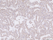
Microscopically, the tumor revealed a well-circumscribed tumor composed of oval to spindle cells concentrically arranged around numerous varying-sized vessels (Fig.3,4). Degenerative nuclear atypia, hemosiderin laden macrophages, and stromal myxoid change are seen (Fig.5,6). There is no increased mitotic activity nor tumor necrosis. Surgical margin is free of tumor. The immunohistochemical (IHC) staining of the resected specimen revealed the tumor cells are diffusely positive for SMA (Fig.7), h-Caldesmon (Fig.8) and partially reactive for CD34; while, they were negative for desmin, HMB-45, Melan-A, CD117, PAX8 and P16. Ki67 proliferation index was up to 3%. The results are consistent with the diagnosis of renal myopericytoma. The patient is alive and free of disease at 4 months after diagnosis.
[Discussion and Conclusion]
Myopericytoma is an uncommon mesenchymal neoplasm and is one of the members of the perivascular tumor family, which includes myofibroma, angioleiomyoma, glomus tumor, myopericytoma, and lesions with hybrid features (such as glomangiopericytoma). Myopericytomas are typically found in the skin and superficial soft tissues, most commonly in the extremities or occasionally in the head and neck or trunk. Rarely, these tumors have been reported to occur in visceral organ, including thorax, heart and brain, and only few cases in kidney had been published. Myopericytomas can arise at any age, however, most are seen in adults with a male predominance. The latest WHO classification of kidney tumors does not list pericytic tumors since those tumors were exceptionally rare in the kidney. There is no standard treatment of renal myopericytoma, while the ten cases had been published were all undergone partial or radical nephrectomy and without recurrence of disease during regular follow up. Myopericytoma is generally considered a benign lesion, although local recurrence and malignant change has been demonstrated in a small portion of cases in soft tissue and skin. It is still unclear that myopericytomas occurring in visceral organs such as the kidney will behave in a similar fashion with this tumor observed at soft tissue sites. In conclusion, this is the first case of renal myopericytoma had been reported in Taiwan who undergone partial nephrectomy and free of disease at 4 months after diagnosis.
Fig.1 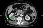 Fig.3
Fig.3 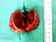 . Fig.5
 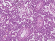 . Fig.7
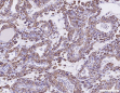
Fig.2 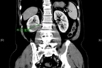 . Fig.4
 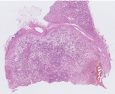 Fig.6
Fig.6 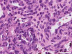 . Fig.8