遠端輸尿管結石造成腎盂破裂
陳怡璇、阮雍順
高雄市立大同醫院
Spontaneous renal pelvis rupture caused by distal ureteral stone
Chen Yi-Hsuan, Yung-Shun Juan
Department of Urology, Kaohsiung Municipal Ta-Tung Hospital, Kaohsiung, Taiwan
Case description:
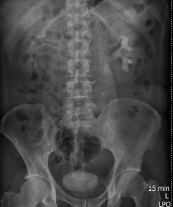
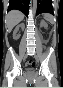
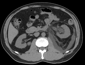
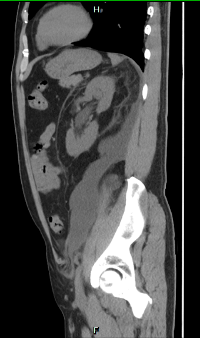
This is a 58-year-old male presented with left flank pain for days. KUB and intravenous pyelography revealed left upper ureteral stone(0.8cm) with left hydronephrosis. (Figure 1). Lab data showed no leukocytosis, but acute kidney injury(creatinine 1.86mg/dl, baseline: 0.8mg/dl). Extracorporeal shock wave lithotripsy was done, but KUB follow up still showed left hydronephrosis and left ureteral stone migration to distal ureter. Due to persistent left flank pain, surgical intervention was suggested. Abdominal CT was arranged and showed left distal ureteral stone with left hydronephrosis(figure 2), fluid accumulation over retroperitoneum(figure 3 and 4), suspected renal pelvis spontaneous rupture. Left ureterorenoscopic lithotripsy and double J insertion were performed smoothly, and the symptoms relieved after operation. There was no complication after the operation and he recovered well upon outpatient department follow up.
Discussion:
Renal pelvis rupture is a rare complication of obstructive uropathy, most happened in the patients with distal ureter or ureterovesical junction stone. The symptoms are similar to renal colic: flank pain, nausea, vomiting. It is hard to differentiated renal pelvis rupture by physical exam, X-ray and echo. Abdominal CT can be the best tool for diagnosis, which would showed peri-renal and retroperitoneum fluid accumulation and extravasation of the contrast medium in delayed phase. The treatment is to release the intra- renal pelvis pressure, including percutaneous nephrostomy or endoscopic surgery.
References:
- Porfyris O, Apostolidi E, Mpampali A, Kalomoiris P. Spontaneous rupture of renal pelvis as a rare complication of ureteral lithiasis. Turk J Urol. 2016 Mar;42(1):37-40. doi: 10.5152/tud.2015.92979.
- Tuncay Tas, Basri CakJroglu, and Süleyman Hilmi Aksoy. Spontaneous Renal Pelvis Rupture: Unexpected Complication of Urolithiasis Expected to Passage with Observation Therapy. Hindawi Publishing Corporation Case Reports in Urology Volume 2013
Figure.1 Figure.2
 .
. 
Figure.3 Figure.4
 .
. 