同時性後腹膜神經纖維瘤與惡性周邊神經鞘腫瘤合併右側腎水腫在一位神經纖維瘤症候群第一型的患者-罕見病例報告
徐明蔚、楊啟瑞
中國醫藥大學附設醫院 泌尿部
Concurrent retroperitoneal neurofibroma and malignant peripheral sheath tumor with hydronephrosis in a patient with neurofibromatosis type 1
Ming-Wei Hsu, Chi-Rei Yang
Department of Urology, China Medical University Hospital, Taichung, Taiwan
Malignant peripheral nerve sheath tumors (MPNSTs) are a type of cancer that are highly aggressive and rare in the general population, but occur at a much higher frequency (2%-5%) in patients with neurofibromatosis type 1 (NF1). Patients with NF1 have a significantly higher risk (8%-13%) of developing MPNSTs, which can be life-threatening. MPNSTs tend to occur in deep locations in the body, and it is important to differentiate them from less harmful neurofibromas in patients with NF1. In fact, approximately 40% of MPNSTs are located in deep tissue and concurrent retroperitoneal tumors are rarely reported. We will present a case with concurrent retroperitoneal neurofibroma and malignant peripheral sheath tumor who presented with hydronephrosis initially and treated successfully by surgical intervention.
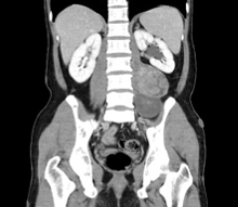
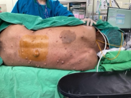
A 42-year-old man with a history of neurofibromatosis type 1 presented to a clinic with persistent nausea and vomiting for days and the abdomen ultrasound showed left renal hydronephrosis. Later abdomen CT scan revealed two left retroperitoneal tumors that were situated close to each other. The upper one was compressing the ureter, causing left hydronephrosis. (Figure 1) There was a high suspicion that the tumors had invaded the bone, psoas muscle, and nerves. He was then referred to CMUH, where a physical examination showed that he had several cafe au lait spots over his trunk and multiple soft, pea-sized bumps on or under his skin. (Figure 2) A spine MRI was performed, which revealed that the two adjacent left retroperitoneal tumors were arising from L3/L4 and L4/L5 vertebral foramen. This finding favored the possibility of neurofibroma or MPNST.
To confirm the diagnosis, the patient underwent a CT-guided biopsy. The pathology report revealed that he had one benign neurogenic tumor and the other one was a low-grade sarcoma. Left URS showed left L/3 ureteral stricture and tumor compression, which was treated with left JJ stent indwelling. The patient then underwent retroperitoneal tumor excision to remove the tumors.
During the surgery, two adjacent left retroperitoneal tumors measuring 8x8cm and 5x5cm were located along the psoas muscle and arose from the L3/L4 and L4/L5 foramen, respectively. The tumors were found to be adhering to the L2, L3, L4, and femoral nerves, making dissection difficult. Unfortunately, L2 was accidentally transected, but this was later repaired by a neurosurgeon. The tumors were eventually ligated from the spinal foramen and removed en-bloc.
After the surgery, the patient experienced left lower limb weakness, with an initial muscle power of around 3. However, he gradually regained left lower limb muscle power through rehabilitation, and was able to walk with a walker smoothly. Otherwise, his post-operative care was uneventful. During the follow-up at the outpatient department, the patient was able to walk freely.
 .
. 
Figure1 and figure 2
Conclusion
It is uncommon that patients with neurofibromatosis have concurrent retroperitoneal tumors, particularly sarcoma. Accurately diagnosing individual tumors prior to surgery is crucial as it impacts the surgical plan. In some cases, imaging studies may not be conclusive and additional pathology testing may be necessary to confirm the diagnosis.