Podium 01
膀胱血管瘤假象下的殺手:原位癌的詭異模仿
唐慈翊1,2、李永進1,2
高雄市立小港醫院1泌尿科
高雄醫學大學附設醫院2泌尿科
Carcinoma in situ of the bladder masquerading as bladder hemangioma
Tsz-Yi Tang1,2, Yung-Chin Lee1,2
1Department of Urology, Kaohsiung Municipal SiaoGang Hospital, Kaohsiung, Taiwan;
2Department of Urology, Kaohsiung Medical University Hospital, Kaohsiung, Taiwan
Case report
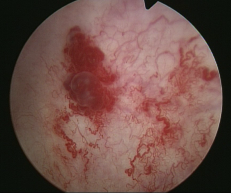
This 71-year-old male patient had a medical history of hypertension He was previously diagnosed with bladder cancer, pTa, and had undergone laser transurethral resection of bladder tumor on 26 July 2022. He reported experiencing off-and-on hematuria for several days. Physical examination revealed a soft abdomen. Urinalysis showed microscopic hematuria without pyuria. Urine cytology showed negative. Cystoscopy on 1 March 2022 revealed a vascular mass at the posterior wall of the bladder (Figure 1). Transurethral resection of bladder tumor was done. Pathology results revealed urothelial carcinoma in situ. The patient was discharged one day after the surgery. The patient was initially diagnosed with a bladder hemangioma based on imaging and cystoscopic findings. However, upon histopathological examination of the biopsy sample, carcinoma in situ was identified.
Carcinoma in situ is a pre-cancerous condition where abnormal cells are found in the epithelial lining of the bladder. Diagnosis is typically made through biopsy, where a tissue sample is taken from the affected area and examined under a microscope. If left untreated, carcinoma in situ may progress to invasive cancer, which can be more difficult to treat and may have a poorer prognosis. Therefore, early detection and prompt treatment are important for improving outcomes. Regular monitoring and follow-up care may also be necessary to monitor for any signs of recurrence.
Conclusion:
we report a case of carcinoma in situ mimicking bladder hemangioma. CIS of the bladder is a high-grade, flat lesion restricted to the urothelial layer. Histopathological and molecular investigations demonstrate that CIS shares morphometric similarities with muscle-invasive bladder cancer (MIBC). Due to the fact that CIS can range from normal-appearing mucosa to a lesion indistinguishable from an inflammatory condition, a pathology report is necessary for the diagnosis. Yet, it has been demonstrated that the introduction of new optical technology improves detection.
Figure 1, Cystoscopy showing an extensive sessile and partially pedunculated vascular mass