Podium 01
膀胱內的爆米花:囊性併腺性膀胱炎
唐慈翊1,2、李永進1,2
高雄市立小港醫院1泌尿科
高雄醫學大學附設醫院2泌尿科
The 'Popcorn in the Bladder': cystitis cystica et glandularis
Tsz-Yi Tang1,2, Yung-Chin Lee1,2
1Department of Urology, Kaohsiung Municipal SiaoGang Hospital, Kaohsiung, Taiwan
2Department of Urology, Kaohsiung Medical University Hospital, Kaohsiung, Taiwan
Case report
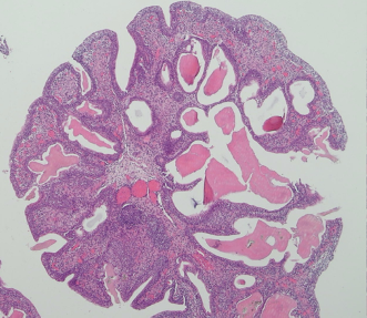
A 48-year-old woman visited the urology clinic due to incidental microscopic hematuria discovered during a routine health examination. Bladder ultrasonography revealed a small bladder tumor, while an abdominal computed tomography (CT) scan with intravenous contrast identified focal thickening of the bladder wall, indicative of a bladder tumor with cT2 staging. Urine cytology results were negative. Upon cystoscopy, a prominent bladder tumor was observed at the posterior wall of the bladder, seemingly connected to a single supplying vessel (Figure 1). En-bloc laser resection of the bladder tumor was performed, and a subsequent pathological examination confirmed Cystitis cystica et glandularis (CCEG) – a benign bladder mucosal lesion characterized by von Brunn's nests (Figure 2), which lead to cystically dilated cystitis cystica and metaplastic cystitis glandularis within the urinary bladder epithelium. Although the precise pathophysiology of CCEG remains unknown, chronic irritation of the bladder mucosa is a probable contributing factor. CCEG is typically asymptomatic, and lesions are often detected incidentally during cystoscopy. The patient was discharged one day post-surgery, and during the one-year follow-up, no recurrence was detected on cystoscopy.
Conclusion:
We report a case of carcinoma in situ (CIS) mimicking bladder hemangioma. CIS of the bladder is a high-grade, flat lesion confined to the urothelial layer. Histopathological and molecular investigations reveal that CIS exhibits morphometric similarities to muscle-invasive bladder cancer (MIBC). Given that CIS can range from normal-appearing mucosa to a lesion indistinguishable from an inflammatory condition, a pathology report is essential for diagnosis. Nevertheless, studies have shown that the incorporation of advanced optical technology enhances detection.
Figure 1, Cystoscopy showing an extensive sessile and partially pedunculated vascular mass
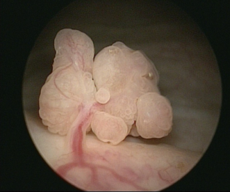
Figure 2, pathological finding showing the presence of Brunn nests, which are invaginations of the urothelium into the lamina propria (20X)